There's a Black Spot on My Tooth — A Dobong Station Dentist's A-to-Z on Telling Decay, Stain and Tartar Apart
Key takeaway A dark spot on a tooth could be decay, coffee or tobacco staining, tartar, or discolouration around an old filling. The eye alone cannot tell them apart — position, texture and progression do. The chalky white spot that signals early decay, how the examination works, treatment and cost by identity, and prevention.
Hello. I am Dr. Oh Ji-hwan of Yonsei The Strong Dental Clinic.
There are things you suddenly notice while brushing under the bathroom light: a thin dark line in the grooves of a molar, a dusky shadow between front teeth, a brownish band near the gums. From the day you spot it, your tongue keeps returning to that place. A patient from Ui-dong showed me a zoomed-in phone photo and asked, "This is a cavity, right?" — while an office worker from near Dobong Station said the opposite: "I assumed it was coffee staining and left it for a year." The interesting part: both guesses were wrong. The first was staining; the second was decay.
The conclusion of today's article first: a dark spot on a tooth has several possible identities, and the eye cannot tell them apart. It may be decay, food or tobacco staining, tartar, or discolouration around an old restoration. And one more thing many people don't know — the first sign of decay is often not black but a chalk-white spot. Today we will line up the suspects one by one, see how the clinic identifies them, what treatment and cost each identity carries, and cover the prevention that matters more than the removal.
The suspects — four in the line-up
The main suspects behind a dark mark on a tooth are four.
First, decay. Acid produced by bacteria dissolves the tooth's minerals. Early on it shows white (demineralisation); as it advances it turns brown, then black. The narrow, deep grooves (fissures) on molar chewing surfaces are decay's favourite starting point — toothbrush bristles cannot reach their floor — which is why "a black line on the chewing surface" is a consultation staple. The key feature: decay changes structure. The surface roughens, pits, and spreads with time.
Second, extrinsic staining. Pigments from coffee, black tea, wine, curry and tobacco attach to the thin film on the tooth surface. The key feature: it sits on the surface while the structure stays sound. It tends to spread across several teeth and often appears as a band along the gumline. Scaling and polishing remove it, and removal leaves the tooth unharmed.
Third, tartar. Plaque hardened with minerals from saliva; old tartar below the gumline can look black. It feels like a hard deposit at or under the gum margin — and it is not a cosmetic matter but a cause of gum disease, so it is removed on sight.
Fourth, restoration-related discolouration. Enamel around an old amalgam can show dark from the metal, or the margin of a resin filling can open and let stain seep into the gap. The latter may be the doorway to secondary decay, not mere staining, and needs checking. We covered this fully in the old filling replacement guide.
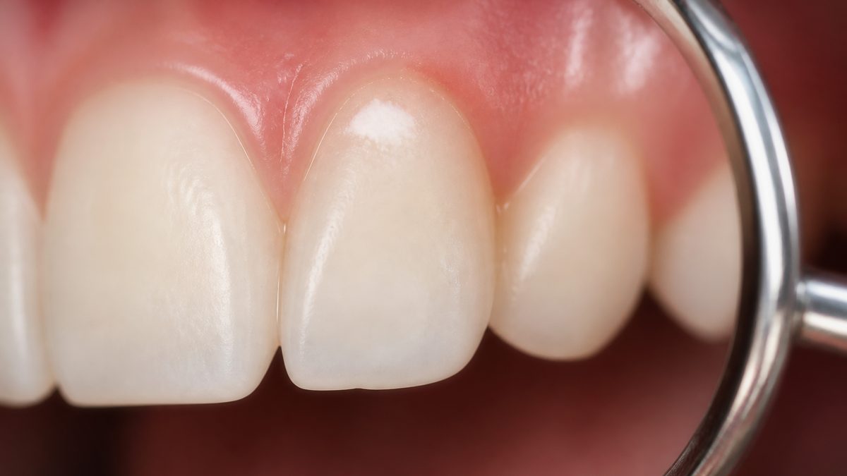
And a fifth, off-list suspect: the white spot. If you went looking for a black dot and found a white patch instead, pay it more attention, not less. A matt, chalk-like white spot can be early demineralisation — minerals beginning to leave the enamel; the opening signal of decay. The good news at this stage: no hole has formed yet, so there is room to reverse it with fluoride and care rather than drilling. I have written in a Ziksir article (in Korean) about what happens when decay is left to grow; this stage is the opposite end of that story — the cheapest point at which to win.
Suspects compared
| Category | Decay | Extrinsic stain | Tartar | Restoration discolouration | Early demineralisation (white spot) |
|---|---|---|---|---|---|
| Colour | White → brown → black | Brown, black | Yellowish; black below gum | Grey, dark rim | Chalky white |
| Typical site | Fissures, between teeth, gumline | Several tooth surfaces, band along gumline | Gum margin, behind lower front teeth | Around an old filling | Near gumline, around braces |
| Surface feel | Rough or pitted | Smooth | Hard deposit | Step or gap | Matt, cloudy |
| Progression | Widens and deepens | Little change | Builds up slowly | Gap can open | Reversible with care |
| Response | Treatment by size | Scaling and polishing | Scaling | Assess, then decide on replacement | Fluoride and observation |
As the table shows, colour alone gives no verdict. Site, surface and progression must be read together — and surface and progression are hard to judge in a bathroom mirror. Treat this table not as a self-diagnosis tool but as pre-reading for the explanation you will hear in the clinic.
Signals that make checking urgent
- The spot's location is sensitive to cold or sweets
- Your tongue or floss catches or shreds there
- The spot has grown noticeably within months
- Chewing on that area is uncomfortable or gives a twinge
- The gum around the spot swells or bleeds
- The margin of an old filling has darkened
- A white patch looks matt and cloudy, without lustre
Any one of these tips the odds towards decay or tartar rather than stain. Conversely, even spread across many teeth with no symptoms suggests staining — but again, the final call belongs to the examination.
How the clinic tells them apart
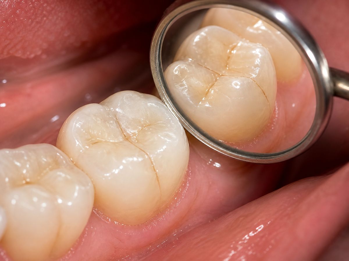
Identifying a dark spot is quicker than you expect, and painless.
- History. Since when, has it grown, has it ever been sensitive, what are your coffee and tobacco habits. These few answers halve the suspect list.
- Inspection and touch. Bright light and magnification on the surface; a blunt-tipped instrument tests texture. Smooth points to stain; catching or softness points to decay.
- X-ray. The visible dot can be the tip of the iceberg. Decay between teeth and secondary decay beneath fillings show only on film. A small surface mark can be broad underneath — this step is not skipped.
- Progression call. If it is decay, we sort "treat now" from "watch." A shallow, arrested lesion can be managed and observed. Drilling everything on sight is not the answer.
- Treatment by identity. Stain — scaling and polish; tartar — removal; decay — treatment by size; demineralisation — fluoride and a care plan. Each branch is explained, and we decide together.
Questions we are often asked
Q. Will whitening remove the black spot? A. Depends on the identity. Surface stain can brighten with whitening and polishing, but decay does not bleach away — sensitivity may only worsen. That is why decay and tartar are ruled out before whitening. For whitening itself, see our whitening guide.
Q. My child has a black line in a molar groove. A. Children's molar fissures are narrow and deep — decay's favourite starting point. We check whether the line is shallow stain or active decay, and if the grooves run deep, we discuss sealing them preventively. Sealants are covered in the sealant and fluoride guide.
Q. I smoke, and I keep second-guessing stain versus decay. A. Tobacco stain tends to spread broadly on the inner surfaces of many teeth. But smokers also carry higher gum-disease risk, so pairing the stain check with a gum examination makes the visit worthwhile. It would be a shame to leave with only the stain polished.
Q. My floss keeps shredding at one particular spot. A. Remember that spot. It can signal interproximal decay roughening the surface, or an opened filling margin. Recurring at the same place, it needs a look.
Q. Can I scrub the spot off at home with baking soda? A. Not recommended. Scrubbing with an abrasive may fade some stain but grinds enamel with it. Stain removal done in the clinic, by methods that spare the tooth, serves the tooth better in the end.
Treatment and cost by identity — what insurance covers
| Identity | Treatment | Insurance | Out-of-pocket guide |
|---|---|---|---|
| Extrinsic stain | Scaling and polishing | Once-yearly scaling covered | About KRW 10,000–20,000 |
| Tartar | Scaling, gum treatment | Covered | About KRW 10,000–50,000 |
| Early demineralisation | Fluoride, care plan | Conditionally covered | A few thousand – tens of thousands |
| Shallow decay | Resin filling | Conditional (children's molars etc.) | About KRW 80,000–200,000 if uncovered |
| Moderate decay | Inlay | Not covered | About KRW 300,000–500,000 |
| Deep decay | Root canal + crown | Root canal covered | RCT ~KRW 50,000–150,000 + crown ~300,000–600,000 |
| Secondary decay at a filling | Remove old filling, restore | Varies by item | Depends on extent |
These figures are orientation ranges and vary with site, size, material and co-payment rate. The one thing to read from this table: solve it in the top-left (stain, demineralisation) and you pay in the tens of thousands; let it reach the bottom-right (deep decay) and you pay in the hundreds of thousands. The same dark spot — the timing of the check decides the number of digits.
Cost-saving checkpoints
- Use the once-yearly insured scaling. Stain and tartar go together, and hidden decay is often discovered in the process.
- Do not miss the white-spot stage — the only stage with room to reverse without drilling.
- Ask to have old filling margins reviewed at check-ups. Early secondary decay can end in a partial repair.
- Check before sensitivity begins. Symptoms usually arrive after the lesion has grown.
After removal — keeping it from coming back

After the spot is removed or treated, keeping the same place from darkening again differs a little by identity.
If you stain easily
- Rinsing with water after coffee, tea or wine alone changes the pace of staining.
- A straw for dark drinks reduces contact with front-tooth surfaces.
- Tobacco is the strongest staining agent and a gum-disease risk in one. Every reduction repays honestly.
- The few days right after stain removal are when colour reattaches easily — rest the dark foods briefly.
If you have had decay or demineralisation
- Use fluoride toothpaste and rinse lightly after brushing so fluoride lingers on the surface.
- Sipping sweet drinks slowly over a long time is the most damaging habit — dwell time in the mouth matters more than total volume.
- If the lesion was between teeth, floss and interdental brushes are the core of prevention — methods in the floss and interdental brush guide.
- If you have been told your fissures run deep, ask about sealants.
For everyone
- Comparing with previous photos and X-rays at 6–12-month check-ups is the surest way to catch change.
- When you find a new dark spot, photograph it and note the date. "Has it grown?" is the single most valuable piece of diagnostic information.
Move your appointment up if
- Pain or sensitivity newly appears at the spot
- The spot grows or the surface looks pitted
- An old filling's margin suddenly darkens
- Floss shreds repeatedly at the same place
Where to have it checked
- Do they identify before they remove? A clinic that examines and explains first beats one that reaches straight for a blanket treatment.
- Can they say "let's watch it"? A clinic that offers observation for an arrested shallow lesion can also be trusted when it says drilling is needed.
- Do they show you on photos and film? Seeing the site and size yourself makes next visit's comparison meaningful.
- Do they talk prevention? Guidance that stops recurrence is worth more, long-term, than removal technique.
- Is it easy to keep visiting? Half of stain and decay management is check-up attendance.
Three common failure patterns
First, spending a year on "it's probably just stain." Like the patient in the opening, a spot believed to be stain sometimes is decay. Checking takes one visit.
Second, fearing "it must be a cavity" and postponing. The failure in the opposite direction. Worry does not arrest a lesion. If it turns out to be stain, the worry ends that day; if it is decay, you caught it at its smallest. Either way, checking wins.
Third, removing and re-removing without changing habits. If habits stay the same after stain removal, the same spot darkens again within months. Changing one prevention habit on removal day is the real treatment.
In closing
Three things, in summary. First, a dark spot can be decay, stain, tartar or restoration discolouration — and colour alone cannot separate them. Site, surface and progression must be read together, and half of that reading needs an examination. Second, decay's first signal is often a white spot, not a black one — and that stage is the only window that can be reversed without drilling. Third, solved at the stain/demineralisation stage the cost sits in the tens of thousands of won; grown into deep decay it climbs into the hundreds of thousands — when you check decides the digits. If you have wondered for weeks what that spot in the mirror is, the answer lies in an examination, not on the tip of your tongue. Individual cases differ, so the precise call comes after we look.
Today in one line: a dark spot on a tooth cannot be identified by eye. If it's stain, the worry ends today; if it's decay, you caught it at its smallest — either way, checking wins.
Our clinic sees patients until 9 p.m. on weekdays and opens 365 days a year, so a worry spotted on the way to work need not be postponed. Our clinic's AI phone guidance also runs 24 hours a day after closing. We are about a five-minute walk from Exit 1 of Dobong Station, Seoul (towards Dobong 1-dong Post Office), an easy bus ride from Ui-dong. For your first visit, see the directions page; if driving, the parking guide.